
Vascular Imaging
What is Vascular Imaging?
Vascular imaging is a medical imaging technique used to visualize and diagnose conditions affecting the blood vessels in the body. This can include the arterial and venous systems, including the heart and brain. Vascular imaging techniques that are available at Innovative Medical Imaging are Echocardiography, Vascular Sonography, and Arterial Ultrasound. These techniques can provide detailed images of the blood vessels and can be used to detect and diagnose a variety of conditions. They can also be used to monitor the progression of vascular diseases, create treatment plans, and evaluate the effectiveness of current treatments.
Non-Invasive Vascular Ultrasound Services we offer:
What are the different Vascular Imaging options?
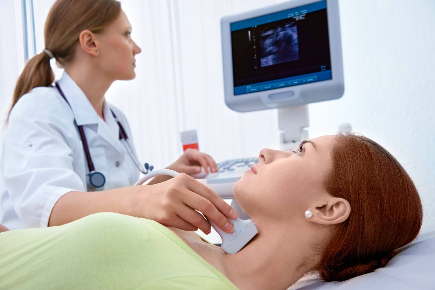
Vascular Sonography
Vascular sonography, also known as vascular ultrasound or duplex ultrasonography, is a medical imaging technique used to produce images of the blood vessels and the blood flow within them. It uses ultrasound to create detailed images of the arteries in different parts of the body. Vascular sonography can be used to evaluate the health of the blood vessels and detect any abnormalities, such as blockages, clots, or aneurysms. It is commonly used to diagnose and monitor conditions such as peripheral artery disease, deep vein thrombosis, and varicose veins. Vascular sonography is a safe, painless, and non-invasive alternative to traditional angiography.
Arterial ultrasound
Arterial ultrasound is a type of vascular sonography that uses ultrasound technology to produce images of the arteries. It is a non-invasive diagnostic technique used to evaluate the health of the arterial system. The ultrasound images are used to assess the blood flow and detect any blockages or aneurysms in the carotid, peripheral vessels, aorta, and renal and mesenteric vessels.
Arterial ultrasound is often used to evaluate peripheral artery disease (PAD) which is a condition in which the blood vessels in the legs and feet become narrowed, limiting blood flow to the lower extremities.
To learn more about vascular imaging contact Innovative Medical Imaging today.
