How does a CT scan work?
A CT scan, or computed tomography, is an imaging procedure that uses X-ray equipment to create a detailed picture of the inside of your body. It is also known as a computerized axial tomography (CAT).
The images built during a CT scan record the body in 2-D “slices” which can then be viewed on their own or layered together to build a 3-D image. Computer software creates both of these image types. Current CT machines build their images in a continuous spiral (or helical) rather than traditional “layers.” This technique is quicker and results in higher quality 3-D images, which can help identify small abnormalities easier than a traditional CT machine.
What is a CT Scan Used for?
Circulatory conditions or diseases:
A CT scan may be used to diagnose blood diseases and conditions such as blood vessel aneurysms and coronary artery disease (atherosclerosis).
Muscular and skeletal injuries:
A CT is helpful for locating and diagnosing spinal conditions, skeletal injuries, head injuries, and internal organ damage.
Additional diseases and conditions:
Some forms of cancer, diseases such as inflammatory bowel disease and sinusitis, and urinary issues such as bladder and kidney stones may be detected and diagnosed with a CT scan.
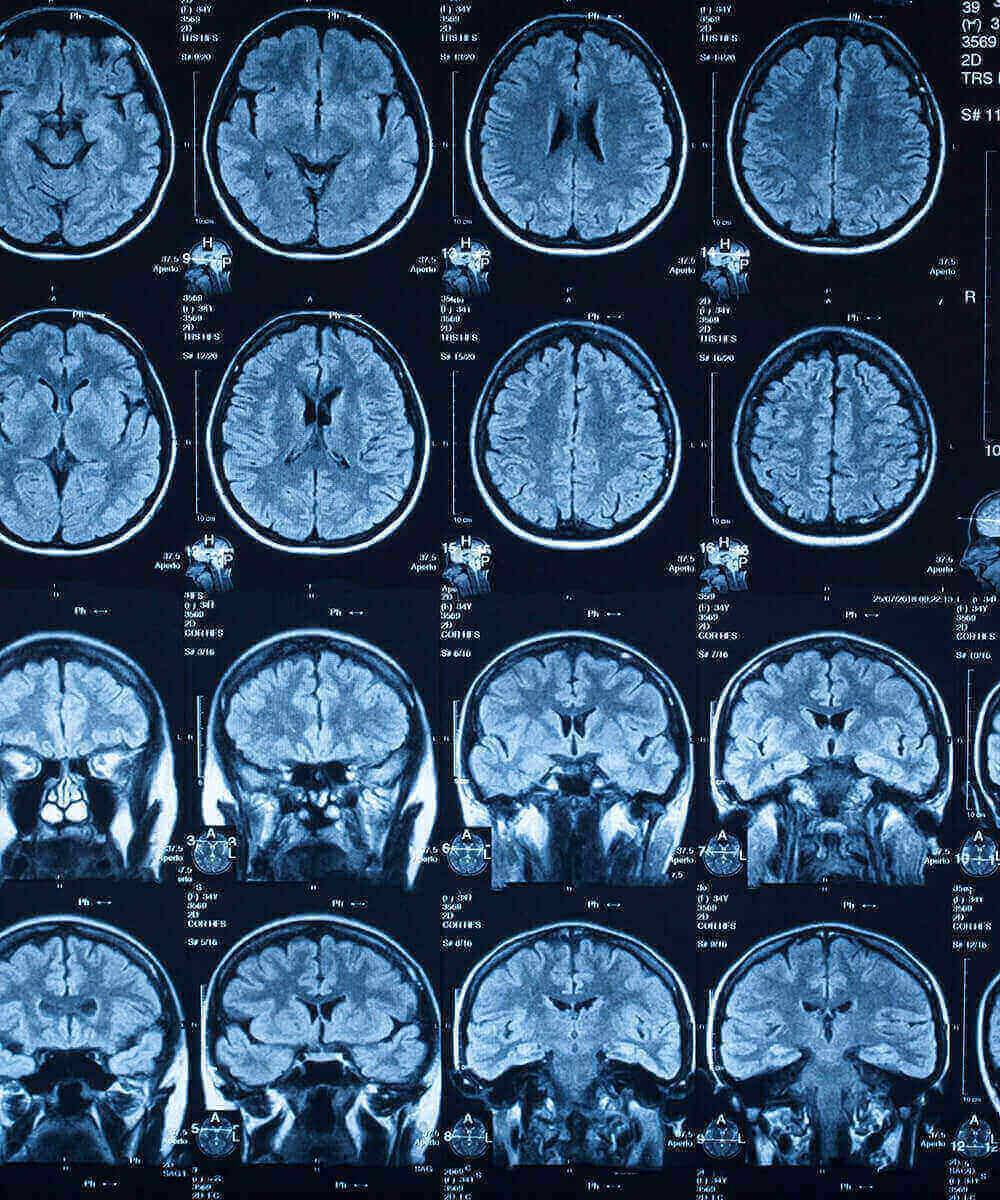
Cognitive diseases and disorders:
CT scans may be used to locate structural issues in the brain like atrophy, hydrocephalus, strokes, and cognitive diseases like Alzheimer’s disease.