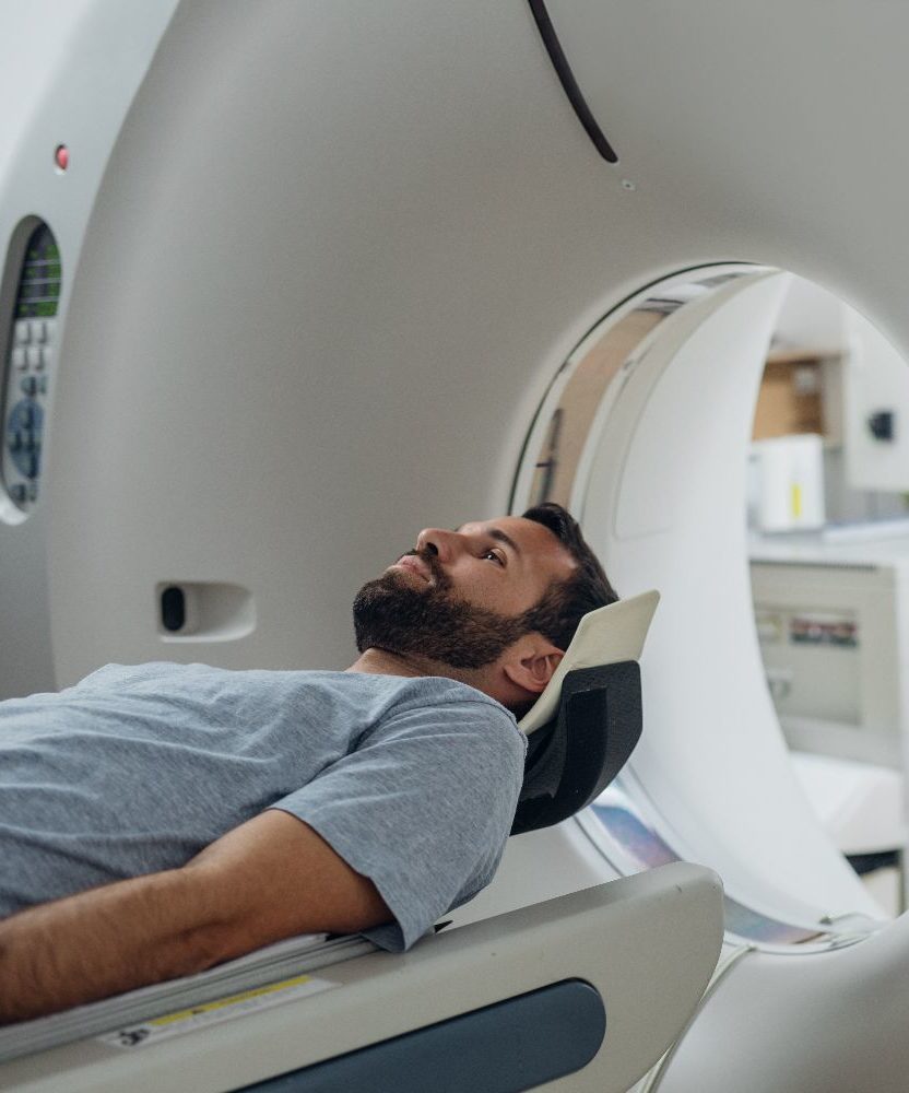
What is an MRI?
Magnetic Resonance Imaging, or MRI, is a non-invasive imaging technology that uses powerful magnets in order to create very detailed 3-D images of body tissues. These powerful magnets cause the alignment of protons within the body tissue, and then a radio frequency current moves the protons out of this alignment. When the radio frequency current stops, MRI sensors detect the alignment changes as the protons realign themselves.
Contrast agents may be given via an IV in order to increase the proton alignment in the magnetic field. The quicker the protons align the brighter the final image appears.
What it used for?
An MRI can be used for:
Soft tissues:
MRIs are good at building images of the soft tissues — they provide clearer images than X-rays or CT scans provide for tendons, muscles, nerves, ligaments, the spinal cord, and the brain.
Brain scans:
An MRI distinguishes between grey and white brain matter and therefore is often used to diagnose tumors and aneurysms. They are better than CT scans at diagnosing brain atrophy or small strokes.
Frequent or repeat scans:
Different than CT scans or X-rays, MRIs do not use radiation to build images. Thus, they are commonly the type of scan utilized to perform repeat imaging.